Polenta cooking
Polenta is traditionally a slowly cooked dish, sometimes taking an hour or longer to cook. When boiled, polenta has smooth creamy textures, caused by starch gelatinization. During gelatinization starch granules first swell as they absorb water. We have used conventional NMR relaxometry (CPMG-T2, IR-T1) and diffusiometry (PGSE) techniques to measure T2, T1 and ADC of polenta at different cooking times. In addition to that 3D high resolution MR microscopy was employed to obtain proton density images, series of T2 weighted images to calculate T2 map images, and diffusion weighted images to calculate diffusion coefficient images of regular polenta and of instant polenta at different cooking times.
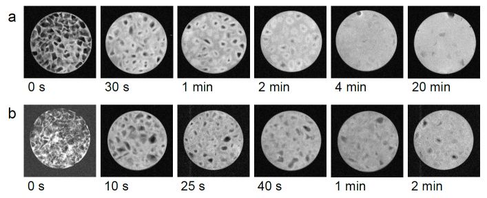
High resolution MR images of regular (a) and instant (b) polenta at different cooking times. Samples were prepared in 8 mm glass tubes by the quenching procedure. The images were acquired by the 3D spin-echo technique at TE/TR=10/2000 ms and slice thickness 0.5 mm; single slices are shown.
Bread baking
The consumer quality of baked products is closely related with dough structure properties. These are developed during dough fermentation and finalized during its baking. In this study, magnetic resonance microscopy (MRM) was employed in a study of dough fermentation and baking. A small hot air oven was installed inside a 2.35-T horizontal bore superconducting magnet. Four different samples of commercial bread mixes for home baking were used to prepare small samples of dough that were inserted in the oven and allowed to rise at 33°C for 112 min; this was followed by baking at 180°C for 49 min. The entire process was followed by dynamic T1-weighted 3D magnetic resonance imaging with 7 min of temporal resolution and 0.23×0.23×1.5 mm3 of spatial resolution. Acquired images were analysed to determine time courses of dough pore distribution, dough volume and bread crust thickness. Image analysis showed that both the number of dough pores and the normalized dough volume increased in a sigmoid-like fashion during fermentation and decreased during baking due to the bread crust formation. The presented magnetic resonance method was found to be efficient in analysis of dough structure properties and in discrimination between different dough types.
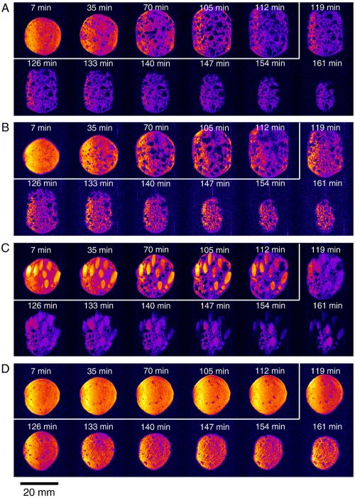
Representative dynamically acquired T1-weighted central-slice MRM images of the bread-making process for mixed flour (A), white flour (B) and seeded flour (C) bread mix samples and for the control (nonyeasted) sample (D). The dough volume increase during fermentation is associated with the yeast activity (7–112 min; enclosed with white lines), while the MRM signal attenuation during baking is associated with the bread crust formation (119–161 min).
Soaking and cooking of the common bean
Legumes can be dried and stored for a very long time if kept in a cool and dry environment. Most of the legumes used for human food require hydration before cooking. The hydration of seeds before or during cooking is essential for protein denaturation, starch gelatinization and seed softening.
Two complementary 3D MRI methods, RARE and SPI, together with T1 and T2 mapping techniques were used for tracking water hydration during the soaking and cooking of common bean seed. The RARE method enabled the detection of free water and excelled in imaging resolution and signal acquisition speed; while the SPI method enabled the detection of bound water and had relatively low resolution and slow signal acquisition. By combining information from the two imaging methods it was possible to track the mobile and bound water that penetrated the bean seed and to determine the hydration dynamics of different anatomical parts of the seed. The study also demonstrated the great potential of MRI to study water imbibition processes in various seeds as also in many other water susceptible materials.
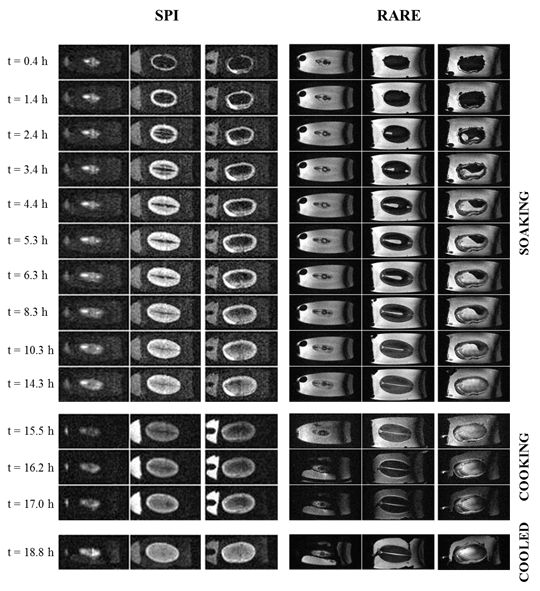
Time series of 3D SPI and 3D RARE images of a bean seed during the 15-hour soaking at room temperature followed by 1.5-hour cooking in near-boiling water; the last set of images was acquired when the seed was cooled back to room temperature. The images are shown in identical three representative slices across the bean seed in a longitudinal orientation; first two are in an orientation perpendicular to the cotyledons (one across the hilum and the other across the center of the seed) and the third is in orientation parallel to the cotyledons.
References:
- SERŠA, Igor, SEPE, Ana, MIKAC, Urška. MRI study of polenta
gelatinization during cooking. Magnetic resonance in food science:
from molecules to man Cambridge: RSC Pub., cop. 2007, p. 141-147.
[PDF
 ]
] - BAJD, Franci, SERŠA, Igor. Continuous monitoring of dough
fermentation and bread baking by magnetic resonance microscopy.
Magnetic resonance imaging, 2011, vol. 29, p. 434-442.
[PDF]
- MIKAC, Urška, SEPE, Ana, SERŠA, Igor. MR microscopy for noninvasive
detection of water distribution during soaking and cooking in the
common bean. Magnetic resonance imaging, 2015, vol. 33, p. 336-345.
[PDF]
- BAJD, Franci, ŠKRLEP, Martin, ČANDEK-POTOKAR, Marjeta, VIDMAR,
Jernej, SERŠA, Igor. Use of multiparametric magnetic resonance microscopy
for discrimination among different processing protocols and anatomical
positions of Slovenian dry-cured hams. Food chemistry, 2016,
vol. 197, p. 1093-1101.
[PDF]
- KRANJC, Matej, BAJD, Franci, SERŠA, Igor, BOEVERE, Mark de, MIKLAVČIČ,
Damijan. Electric field distribution in relation to cell membrane
electroporation in potato tuber tissue studied by magnetic resonance
techniques. Innovative food science & emerging technologies,
2016, vol. 37, p. 384-390.
[PDF]
- BAJD, Franci, ŠKRLEP, Martin, ČANDEK-POTOKAR, Marjeta, VIDMAR,
Jernej, SERŠA, Igor. Application of quantitative magnetization transfer
magnetic resonance imaging for characterization of dry-cured hams.
Meat science, 2016, vol. 122, p. 109-118.
[PDF]
- BAJD, Franci, GRADIŠEK, Anton, APIH, Tomaž, SERŠA, Igor. Dry-cured
ham tissue characterization by fast field cycling NMR relaxometry and
quantitative magnetization transfer. Magnetic resonance in chemistry,
2016, vol. 54, no. 10, p. 827-834.
[PDF]
- BAJD, Franci, ŠKRLEP, Martin, ČANDEK-POTOKAR, Marjeta, SERŠA, Igor.
MRI-aided texture analyses of compressed meat products. Journal of
food engineering, 2017, vol. 207, p. 108-118.
[PDF]