Electric Current Density Imaging
Electric Current Density Imaging (CDI) is a new modality of magnetic
resonance imaging that enables electric current distribution imaging in
conductive samples containing water. CDI techniques may be further
divided to: DC-CDI, a technique designed to image direct electric
current density distribution, AC-CDI technique
enabling imaging of alternating electric current distribution of the
frequency up to a few kHz and RF-CDI that operates at the RF Larmor
frequency. The principle of CDI is based on mapping magnetic field
changes caused by electric currents flowing through the sample. Once
maps of the magnetic field changes are obtained, the electric current
density can be calculated from magnetic field change maps
using Ampere's law.
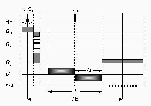
Direct electric current density MR imaging sequence.
CDI sequences utilize electric currents applied in pulses that are well
synchronized with the imaging sequence. Electric pulses produce
temporal shifts of the precession frequency and consequently phase
shifts proportional to the magnetic field change. These shifts can be
added cumulatively by an appropriate arrangement of electric and RF
pulses in the CDI sequence and later detected by a phase sensitive MRI.
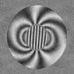
The image of the real MR signal component (left) of the sample
consisting of two concentric cylinders: inner field with saline and
outer with water
(right).
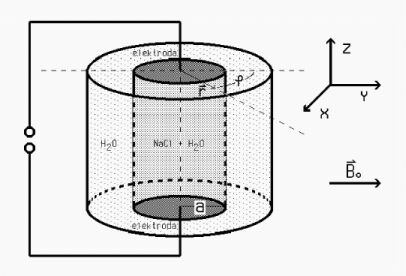
In one sample orientation, only the component of magnetic field change
along the static magnetic field can be measured. Therefore, the sample
has to be rotated in remaining perpendicular spatial orientations to
map other components of magnetic field change. Once this is done the
electric current density can be calculated as the curl of magnetic
field maps.
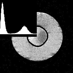
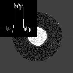
Conventional magnitude MR image (left) and calculated electric current
density image of the same sample (right). The electric current of 20 mA
was flowing through the inner cylinder of the sample.
References:
- SERŠA, Igor, JARH, Orest, DEMŠAR, Franci. Magnetic resonance
microscopy of electric currents. J. magn. reson., Ser. A, 1994,
vol. 111, p. 93-99.
[PDF
 ]
]
- BERAVS, Katarina, WHITE, David, SERŠA, Igor, DEMŠAR, Franci.
Electric current density imaging of bone by MRI. Magn. reson.
imag., 1997, vol. 15, p. 909-915.
[PDF]
- SERŠA, Igor, BERAVS, Katarina, DODD, Nick J. F., ZHAO, Sha, MIKLAVČIČ,
Damijan, DEMŠAR, Franci. Electric current density imaging of mice
tumors. Magn. reson. med., 1997, vol. 37, p. 404-409.
[PDF]
- BERAVS, Katarina, OVEN, Primož, SERŠA, Igor, TORELLI, Niko,
DEMŠAR, Franci. Electric current density imaging of pedunculate oak
(Quercus robur L.) twigs by magnetic resonance imaging. Holzforschung,
1998, vol. 52, p. 541-545.
[PDF]
- MIKAC, Urša, DEMŠAR, Franci, BERAVS, Katarina, SERŠA, Igor.
Magnetic resonance imaging of alternating electric currents.
Magn. reson. imag., 2001, vol. 19, p. 845-856.
[PDF]
- MIKAC, Urša, DEMŠAR, Alojz, SERŠA, Igor, DEMŠAR, Franci. Electric
current density imaging of a tablet dissolution. Cell. Mol. Biol.
Lett., 2002, vol. 7, p. 136-138.
[PDF]
- MIKAC, Urška, DEMŠAR, Alojz, DEMŠAR, Franci, SERŠA, Igor.
A study of tablet dissolution by magnetic resonance electric current density
imaging. J. magn. reson., 2007, vol. 185, p. 103-109.
[PDF]
- SERŠA, Igor. Auxiliary phase encoding in multi spin-echo sequences:
Application to rapid current density imaging.
J. magn. reson., 2008, vol. 190, p. 86-94.
[PDF]
- SERŠA, Igor. Current density imaging sequences with separation of
mobile-ion current from immobile-ion current.
J. magn. reson., 2009, vol. 196, p. 33-38.
[PDF]
- SERŠA, Igor. Enhanced sensitivity current density imaging.
J. magn. reson., 2010, vol. 204, p. 219-224.
[PDF]
- KRANJC, Matej, BAJD, Franci, SERŠA, Igor, MIKLAVČIČ, Damijan.
Magnetic resonance electrical impedance tomography for monitoring electric
field distribution during tissue electroporation. IEEE transactions
on medical imaging, 2011, vol. 30, no. 10, p. 1771-1778.
[PDF]
- ESSONE MEZEME, Melvin, KRANJC, Matej, BAJD, Franci, SERŠA, Igor,
BROSSEAU, Christian, MIKLAVČIČ, Damijan.
Assessing how electroporation affects the effective conductivity tensor
of biological tissues. Appl. phys. lett., 2012, vol. 101, 213702
[PDF]
- KRANJC, Matej, BAJD, Franci, SERŠA, Igor, WOO, Eung Je, MIKLAVČIČ, Damijan.
Ex vivo and in silico feasibility study of monitoring electric field
distribution in tissue during electroporation based treatments.
PloS one, Sep. 2012, vol. 7, no. 9, p. 1-8.
[PDF]
- BAJD, Franci, KRANJC, Matej, MIKLAVČIČ, Damijan, SERŠA, Igor.
Current density imaging during tissue electroporation = Imidľing na gustinata
na struja pri elektroporacija na tkivo. Pril. - Maked. akad. nauk. umet.,
Odd. biol. med. nauki, vol. 33, no. 1, p. 367-372.
[PDF]
- KRANJC, Matej, BAJD, Franci, SERŠA, Igor, MIKLAVČIČ, Damijan.
Magnetic resonance electrical impedance tomography for measuring electrical
conductivity during electroporation. Physiological measurement,
2014, vol. 35, no. 6, p. 985-996.
[PDF]
- KRANJC, Matej, MARKELC, Boštjan, BAJD, Franci, ČEMAŽAR, Maja, SERŠA, Igor,
BLAGUS, Tanja, MIKLAVČIČ, Damijan. In situ monitoring of electric field
distribution in mouse tumor during electroporation. Radiology, 2015,
vol. 274, no. 1, p. 115-123.
[PDF]
- KRANJC, Matej, BAJD, Franci, SERŠA, Igor, BOEVERE, Mark de, MIKLAVČIČ, Damijan.
Electric field distribution in relation to cell membrane electroporation in potato
tuber tissue studied by magnetic resonance techniques.
Innovative food science & emerging technologies, 2016, vol. 37, p. 384-390.
[PDF]
- KRANJC, Matej, KRANJC BREZAR, Simona, BAJD, Franci, SERŠA, Gregor,
SERŠA, Igor, MIKLAVČIČ, Damijan. Predicting irreversible electroporation-induced
tissue damage by means of magnetic resonance electrical impedance tomography.
Scientific reports, 2017, vol. 7, art. no. 10323, p. 1-10
[PDF]
- SERŠA, Igor. Electric current detection by T2* relaxivity change: A Feasibilty Study.
EMBEC 2020, IFMBE proceedings, 2021, vol. 80, p. 470-477.
[PDF]
 Home
Home